点击展开
Gene editing dog model
Gene editing dog model
Backgroundinformation
1 Atherosclerosis
Atherosclerosis (AS) is an elderly multiple disease caused by multiple factors such as heredity and environment. With the continuous improvement of people's living standards, the incidence of atherosclerosis is increasing year by year, and the trend is getting younger. Among them, coronary heart disease and cerebrovascular diseases caused by coronary atherosclerosis and intracranial arteriosclerosis have become the main killers of human beings. Among the many pathogenic factors, lipid metabolism disorder, especially hypercholesterolemia is most closely related to it.
2 ApoE gene
ApoE gene is an important component of plasma lipoprotein, which plays an important role in regulating lipid metabolism and distribution in cells, especially in cholesterol metabolism. Low expression or loss of ApoE leads to an increase in blood lipid levels, which induces atherosclerotic disease. In recent years, ApoE knockout mice have become one of the important animal models for studying the pathogenesis and pharmacology of anti-AS.Although mice AS model has been widely used , due to the long genetic relationship between mice and human beings and their short life span, mice are quite different from human beings in size, physiological structure, biochemical indexes and lipid metabolism pathway, which makes it difficult to accurately simulate the occurrence and treatment process of human diseases. Therefore, there is no animal model to accurately simulate AS disease in related studies.
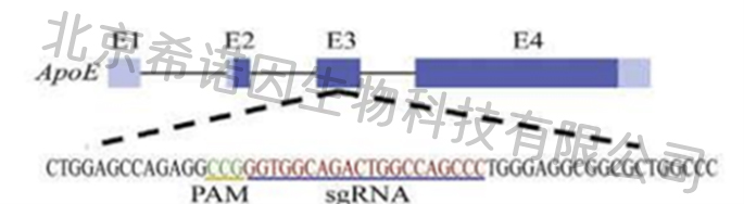
Through whole-genome sequencing analysis of dogs, a total of 19,300 genes were identified, among which 18,000 genes were identical with known human genes, higher than mice and other experimental animals. In addition, dogs are similar to humans in lipid metabolism, physiological anatomy and other aspects, making dogs an ideal model animal for the study of human cardiovascular and cerebrovascular diseases. Therefore, we conducted gene editing on ApoE exon 3, and obtained four F0 generation gene editing dogs: ApoE exon 3 double-allele mutation -34bp/+17bp and -2236bp.
Results of F0 generation of model dogs
1hyperlipidemia
The 4 F0 generation dogs showed hyperlipidemic phenotypehigh with cholesterol and high low-density lipoprotein after birth.
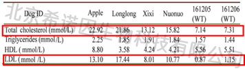
Blood lipid at 30 days after birth (Published data)

Blood lipid at1.5-2 years (Unpublished data)
2Atherosclerosis and complications
Vascular B ultrasound
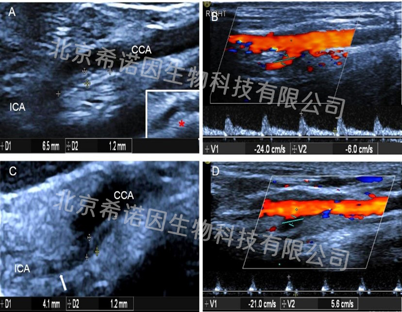
Vascular ultrasound examination was performed at nearly 2 years of the F0 generation. The results showed that 6.5×1.2mmechoic flat plaque was detected in the posterior wall of the initial segment of the right internal carotid artery in ApoE gene edits dogs (FIG. A), and the flow rate was 24/6cm/s (FIG. B), and the flow rate of the internal carotid artery was 20/6cm/s. 4.1×1.2mm plaque was detected in the posterior wall of the initial segment of the left internal carotid artery (FIG. C), and the flow rate was 21/6cm/s, presenting an "oscillating" flow spectrum (FIG. D). The flow rate of the internal carotid artery was 11/6cm/s, presenting an "oscillating" flow spectrum. Echoic flat plaques of 19.9x1.3mm were detected on the posterior wall of abdominal aorta, plaques of different sizes were detected in renal artery, iliac artery and superficial femoral artery, and vessels of different degrees of stenosis were detected.
Internal carotid atherosclerosis (pictures unpublished)
MRI:
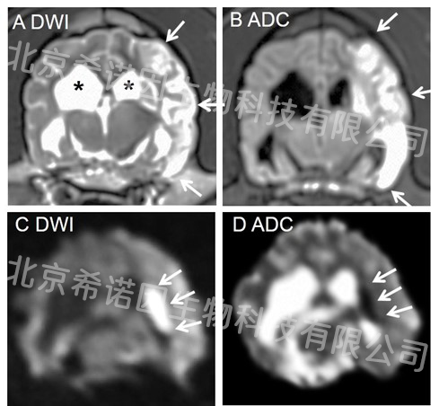
MRI results showed cerebral infarction in ApoE gene editing dogs (Mutant 1). The arrows in Figures A and B show 7.41 ml of massive infarction in the left frontal, parietal, and temporal lobes. The arrows in figures C and D show 1.14 ml of fresh, acute ischemic infarction in the left basal ganglia.
Cerebral infarction (pictures unpublished)
Autopsy results:
1. F0 generation all died of cardiac and cerebrovascular diseases caused by atherosclerosis at about 2 years old. Autopsy showed that myocardial fibrosis and dilation of the heart cavity were caused by coronary artery stenosis and myocardial fiber ischemia and hypoxia.
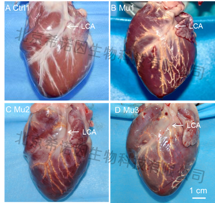
coronary atherosclerosis(pictures unpublished)
LCA left coronary artery.
2. Atherosclerotic plaques were found in both internal carotid arteries and external carotid arteries, and secondary thrombosis was found in left internal carotid artery plaque of Mutant 1 (Figure A).
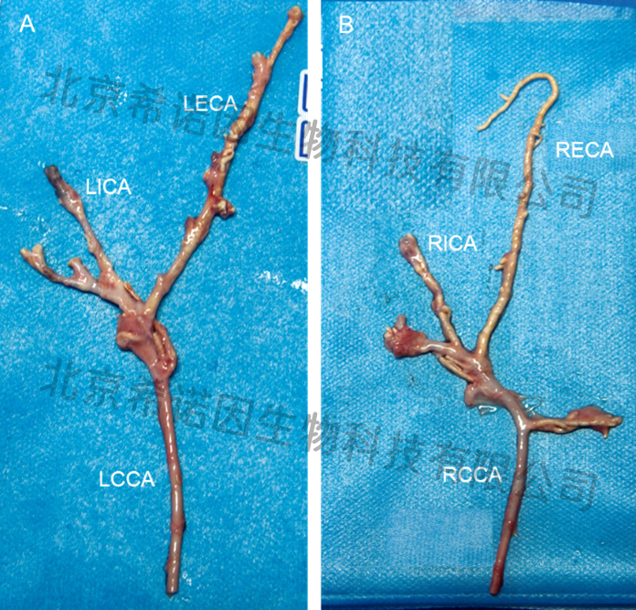
Carotid atherosclerosis (pictures unpublished)
LECA left external carotid artery;LICA left internal carotid artery;LCCA left carotis communis artery.
RECA right external carotid artery;LICA right internal carotid artery;LCCA right carotis communis artery.
3. Atherosclerotic plaques were found in both basilar artery and anterior cerebral artery, secondary thrombosis was found in Mutant 1 basilar artery (Figure B), and the autopsy results were corresponding to the results of preoperative MRI.

Intracranial atherosclerosis (pictures unpublished)
BA basilar artery;RCA rostral cerebral artery.
4. Due to the atherosclerosis of iliac artery, femoral artery and vascular stenosis of the model dog, insufficient blood supply to the hind limb, occlusion of dorsal foot artery and gangrene of the hind limb were generated.
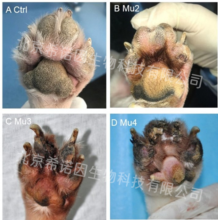
Gangrene (pictures unpublished)
Pathological results:
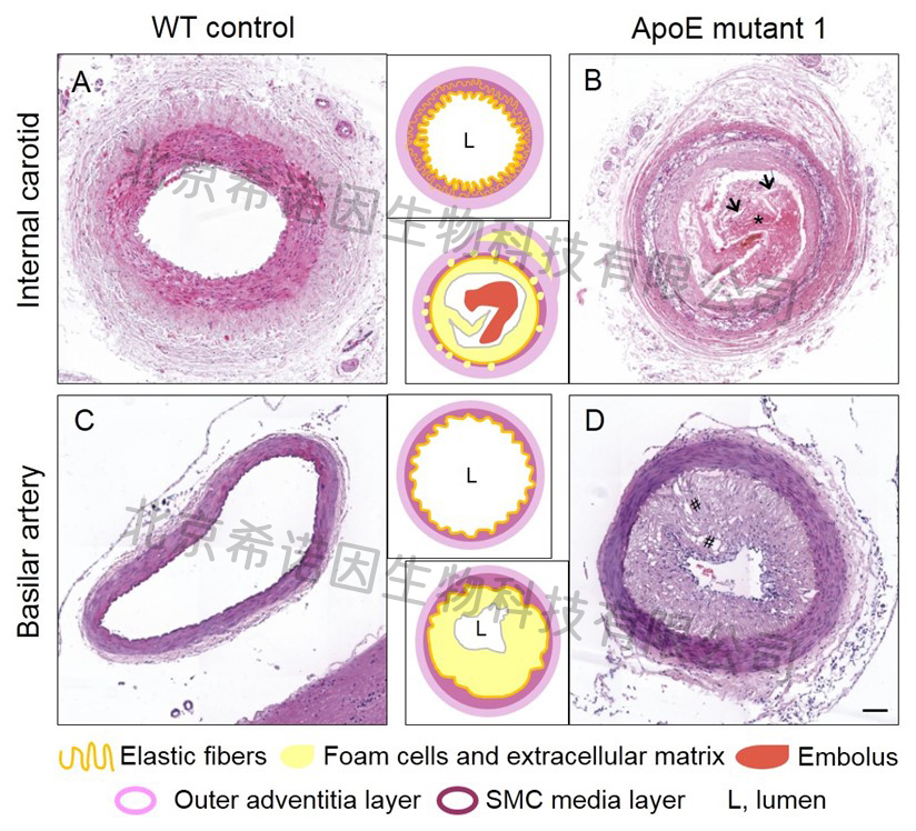
Nervous system diseases are complex diseases occurring in central nervous system, peripheral nervous system and vegetative nervous system with sensory, motor, consciousness and vegetative nervous dysfunction as the main manifestations. After thirty thousand years of convergent evolution and artificial breeding, domestic dogs show rich social, emotion and empathy, and have unique biological characteristics to study neurological diseases. The model of AUTISM spectrum disorder, represented by Shank3 gene editing dogs, show obvious rigid behavior and motor disorders, and the model dogs show obvious A large number of red blood cells (" * "), cholesterol crystals (" → ") and thrombus composed of plaques were found in the lumen of the left internal carotid artery in ApoE model dogs (Figure B). The intima of the basilar artery was significantly thickened, and a large number of foam cells gathered under the intima of the lumen, and there were a large number of small vacuoles in the cytoplasm (" # "), and the lumen was almost occluded (Figure D).
ApoE gene editing dogs always show the hyperlipidemia phenotype of high total cholesterol and high low density lipoprotein after birth. Vascular B-ultra sound can show multiple atherosclerotic plaques in the whole body, such as internal carotid artery, abdominal aorta, renal artery, iliac artery, superficial femoral artery, etc. Most model dogs suffer from late hind limb insufficiency and gangrene. Simvastatin, a clinical lipid-lowering drug, effectively reduced total cholesterol and low density lipoprotein in this model. Autopsy showed tortuous calcification and atherosclerosis of basilar artery and anterior cerebral artery in model dogs, and even thrombosis and cerebral infarction of internal carotid artery and basilar artery in severe cases. The phenotype of ApoE gene edited dogs largely mimics the phenotype of patients with clinical atherosclerosis complicated by cardiovascular and cerebrovascular diseases, and is sensitive to lipid-lowering drug therapy in humans. Therefore, ApoE gene editingdogs are ideal animal models for the study of atherosclerotic related diseases.
3 Population quantity
F0 \ F1, F2 and F3, a total of 120
4Model application
I. Use model dogs to study the pathogenesis of the following aspects:
1. Pathogenesis and mechanism of atherosclerosis;
2. On the basis of ApoE model dogs, the atherosclerosis model dogs with multi-gene editing (such as SR-BI gene, Akt gene, PDGFRβ gene, etc.) were created to further study the pathogenesis and mechanism of atherosclerosis.
3. Pathogenesis and mechanism of atherosclerosis complicated with cardiovascular disease and cerebrovascular disease;
4. Microangiopathic complications such as cognitive impairment and other mechanisms.
II. Use model dogs to conduct drug effectiveness evaluation and drug development experiments:
1. Evaluate the effect of drugs on blood lipid;
2. Evaluate the effect of drugs on plaque formation;
3. Evaluate the therapeutic effect of drugs on atherosclerosis complicated with cardiovascular disease and cerebrovascular disease;
4. Evaluate the therapeutic effect of drugs on atherosclerosis complicated with cognitive impairment, etc.
III. Use model dogs to conduct experiments and evaluations on the effectiveness of non-drug treatments such as medical instruments and surgery;
IV. On the basis of model dogs, animal models of other diseases are induced by dietary induction and surgical operation.


